

If you’re in Yorba Linda and wondering whether to remove your wisdom teeth, this article shows what to expect and how to choose trusted local care. You can get clear answers about whether removal is necessary, what the procedure involves, and how recovery will go, so you can make a confident decision.
The post explains why wisdom teeth often cause problems, common signs that indicate removal, what happens at the consultation, and practical tips for a smooth recovery. It also guides you through choosing a compassionate oral surgeon, payment and insurance considerations, and steps to prevent complications — all in plain, reassuring language.
Expect friendly, expert advice that demystifies the process and focuses on comfort and safety, helping you take the next step with confidence. If questions or concerns linger, the article points toward consultation and practical next steps to get personalized care.
Why Wisdom Teeth Are Removed
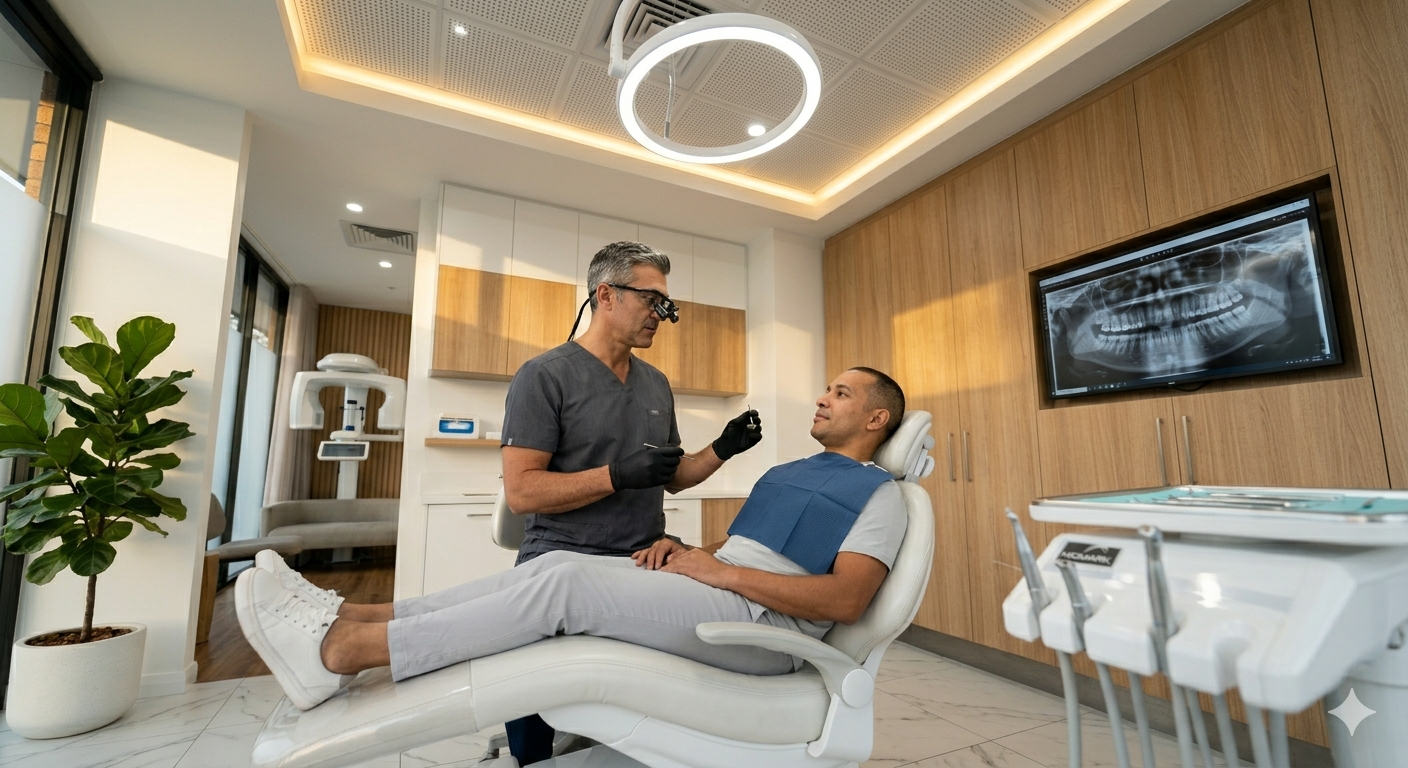
Dentists remove wisdom teeth when those molars threaten oral health, alignment, or comfort. Removal decisions balance current symptoms, X-ray findings, and the risk of future problems.
Common Dental Concerns
Impacted teeth sit trapped under gum or bone and can push against second molars. That pressure often causes crowding, shifts in bite, and damage to adjacent teeth.
Partially erupted wisdom teeth create a flap of gum tissue that traps food and bacteria, leading to recurrent infections called pericoronitis. Pain, swelling, and bad taste are typical signs.
Cavities form more easily on wisdom teeth because they sit far back and are hard to clean. When decay reaches the pulp or weakens surrounding teeth, extraction becomes the safer option.
Orthodontists also recommend removal when wisdom teeth threaten completed braces results or cause arch crowding that compromises long-term alignment.
Potential Risks of Not Removing Wisdom Teeth
Untreated impacted wisdom teeth can form cysts or, rarely, tumors that damage jawbone and nearby teeth. Small cysts may be symptomless at first but enlarge over time, requiring more complex surgery.
Chronic infections around partially erupted teeth increase the risk of spreading bacteria to surrounding tissues or, in people with certain medical conditions, causing systemic complications.
Progressive decay undermines adjacent second molars, sometimes making it necessary to extract otherwise healthy teeth.
For patients with limited mouth access, retained wisdom teeth raise the chance of recurring pain and repeated antibiotic needs, which can complicate long-term management.
Typical Symptoms Leading to Removal
Persistent pain at the back of the mouth that worsens with chewing or radiates to the ear commonly signals a problem. Dentists evaluate pain alongside swelling, redness, and recurrent bad breath.
Repeated infections at the wisdom tooth site—especially pericoronitis episodes within a year—often prompt extraction to stop the cycle.
Visible decay that reaches the root or causes significant bone loss on X-ray usually leaves extraction as the practical choice.
Orthodontic relapse after braces, root resorption of the second molar seen on radiographs, and cyst formation are clear clinical indications for removal rather than continued monitoring.
Signs You May Need Wisdom Teeth Removal
Common indicators include persistent pain in the back of the mouth, repeated gum swelling or infection around the last molars, and noticeable shifts in tooth alignment that affect the bite. These symptoms often prompt dental X-rays and an exam to confirm whether removal will prevent further problems.
Pain and Jaw Discomfort
Persistent pain behind the second molars, especially when chewing or opening the mouth wide, suggests wisdom tooth issues. The pain may be sharp during biting or a dull ache that radiates to the ear or temple.
Pain that worsens over days or recurs frequently often means the tooth is partially erupted or pressing against adjacent teeth. Dentists look for localized tenderness, limited mouth opening (trismus), and pain that doesn’t respond well to over‑the‑counter pain relievers.
If radiographs show impaction or angled growth, removal can relieve pressure and prevent damage to neighboring teeth. He or she will consider the patient’s age, overall health, and pain pattern when recommending extraction.
Swelling or Gum Infections
Red, swollen gums at the back of the mouth that bleed when brushing often indicate pericoronitis, an infection around a partially erupted wisdom tooth. Signs include swollen tissue that traps food, bad taste or odor, and occasional pus drainage.
Repeated or severe infections can spread to adjacent tissues, cause fever, or make swallowing uncomfortable. Dentists typically treat the infection first with cleaning, possible antibiotics, and then evaluate the tooth for extraction to prevent recurrence.
Imaging helps determine if a tooth’s position makes infection likely to return. If so, removal during a controlled setting reduces the risk of complicated infections later.
Changes in Teeth Alignment
Crowding or shifting of nearby teeth—especially the second molars and premolars—can signal pressure from erupting wisdom teeth. Patients may notice newly cramped front teeth, changes in how the upper and lower teeth meet, or orthodontic relapse after braces.
X‑rays reveal whether wisdom teeth push against roots or crowns of adjacent teeth. When crowding threatens restorative work or orthodontic outcomes, dentists often recommend removal to protect long‑term alignment.
Timing matters: younger patients typically heal faster and experience fewer complications. The dental team weighs alignment risk, patient age, and future oral health when advising on extraction.
Call the dental office in Yorba Linda for an exam, X‑rays, and a clear plan tailored to the patient’s symptoms and comfort.
What to Expect During the Consultation
The consultation clarifies the patient’s oral health, treatment options, and timeline. It also identifies medical factors that affect anesthesia and recovery.
Dental Examination and Imaging
The clinician inspects the mouth visually and palpates the gums and jaw to check tooth position and signs of infection. They will assess crowding, gum inflammation, and any nearby teeth at risk.
A panoramic X-ray (OPG) is commonly taken to show all four wisdom teeth, roots, and the lower jaw nerve canal. In cases with complex root anatomy or sinus proximity, a CBCT scan provides a 3D view to guide safer extraction.
The team records bite relationships and may test tooth mobility if decay or periodontal disease is suspected. They also review prior dental records or X-rays if the patient transfers care, ensuring treatment decisions use complete information.
Discussing Treatment Plans
The clinician explains whether each wisdom tooth needs removal now, later, or can be monitored. They use the images and exam findings to justify the recommendation, citing root shape, impaction type (vertical, horizontal, mesioangular, or distoangular), and risk to adjacent teeth.
They outline anesthesia options: local anesthesia, nitrous oxide (laughing gas), oral sedatives, or IV sedation. The discussion includes expected procedure length—typically 20–60 minutes depending on complexity—and whether bone removal or sectioning of the tooth will be necessary.
Risks and benefits receive specific explanation: potential nerve irritation, dry socket rates, infection, and expected pain levels. They also discuss alternatives, such as periodic monitoring or orthodontic referral when relevant.
Personalized Recommendations
The clinician factors in medical history, medications (especially blood thinners), smoking status, and allergies before finalizing a plan. They tailor preoperative instructions, such as fasting for IV sedation or pausing certain medications under physician guidance.
Recovery plans include customized pain control, antibiotic use only when indicated, and step-by-step oral hygiene recommendations to reduce infection risk. The office provides written aftercare, emergency contact numbers, and a follow-up schedule for suture removal or healing checks.
They also give practical logistics: expected time off work or school, transportation needs post-sedation, and costs or insurance estimates. Patients receive clear next steps and are encouraged to ask questions before scheduling.
The Removal Procedure Explained
This section outlines what patients can expect before, during, and after extraction, emphasizing anesthesia choices, the surgical steps, and techniques used to maximize comfort and speed recovery.
Preparation and Anesthesia Options
Patients meet with the dentist or oral surgeon for a focused exam and updated X-rays, usually a panoramic or CBCT, to map tooth position and root anatomy. Medical history, current medications, and any allergies are reviewed to determine safe anesthesia and to plan for bleeding or infection risks.
Local anesthesia is standard for straightforward extractions; a numbing injection blocks pain at the surgical site. For anxious patients or complex impactions, the practice commonly offers nitrous oxide (laughing gas) for mild sedation or oral sedatives taken before the appointment.
IV sedation provides deeper relaxation and is monitored by trained staff; it allows the patient to remain responsive but drowsy. General anesthesia is available for extensive cases or special needs patients and requires fasting and escort arrangements due to longer recovery. The team explains fasting rules, arrival time, and post-op transportation during the pre-op call.
Step-by-Step Overview of Extraction
After anesthesia takes effect, the clinician confirms numbness and positions the patient for clear access and visibility. For erupted wisdom teeth, the dentist loosens the tooth with elevators and removes it with forceps in one piece when possible.
For partially or fully impacted teeth, the surgeon makes a small gum incision and removes any bone blocking access. They may section the tooth into smaller pieces to remove it with minimal bone removal, reducing trauma to surrounding tissue.
Once the tooth is out, the socket is irrigated to remove debris and checked for remaining fragments. The clinician places gauze to control bleeding and may use dissolvable sutures to approximate the gum tissue; these sutures typically dissolve in 1–2 weeks.
Post-op instructions cover pain control, ice application, diet modifications, and activity limits. Clear discharge directions and an emergency contact number ensure prompt help if bleeding, fever, or unusual swelling occurs.
Modern Techniques for Comfort
Minimally invasive techniques, such as piezosurgery or piezoelectric bone cutting, reduce vibration and heat compared with traditional rotary instruments. This precision lowers postoperative swelling and speeds healing for impacted cases.
Local anesthetic formulations often include long-acting agents that keep the area numb for several hours, reducing the need for immediate pain medication. Computer-assisted local delivery and nerve blocks improve accuracy and decrease injection discomfort.
Adjuncts like platelet-rich fibrin (PRF) or collagen plugs promote clot stability and tissue healing when placed into sockets. Many practices offer multimodal pain management protocols—combining acetaminophen, NSAIDs, and targeted opioids only when necessary—to limit opioid exposure.
Continuous monitoring during sedation, use of oxygen, and ACLS-trained staff help maintain safety. These combined techniques focus on reducing anxiety, minimizing tissue trauma, and improving the overall recovery experience for patients.
Recovery and Healing Process
Expect swelling, mild bleeding, and manageable pain after extraction. Following clear aftercare steps, using prescribed medications, and keeping follow-up appointments helps prevent complications and speeds recovery.
Immediate Aftercare Tips
After surgery, the patient should bite gently on gauze for 30–60 minutes to control bleeding. Replace gauze only when bleeding slows; avoid vigorous spitting or rinsing during the first 24 hours to protect the forming clot.
Use an ice pack on the cheek in 20-minute cycles for the first 48 hours to reduce swelling. Keep the head elevated while resting and avoid heavy lifting or exercise for 48–72 hours to minimize bleeding and discomfort.
Follow prescribed medications exactly: take antibiotics to prevent infection and analgesics as directed for pain control. Avoid aspirin and other blood-thinning OTCs unless the dentist approves them.
Do not smoke or use nicotine for at least 72 hours; nicotine delays healing and greatly increases dry socket risk. Eat soft foods like yogurt, mashed potatoes, and smoothies; avoid straws for 7–10 days to protect the socket.
Managing Discomfort at Home
Expect moderate pain that peaks 24–48 hours after extraction and improves over 3–5 days. Use scheduled pain meds during that peak period rather than waiting for severe pain to return faster control.
Apply warm compresses to the jaw after 48 hours to promote circulation and reduce stiffness. Gentle jaw exercises, like opening and closing slowly, help restore normal mobility without stressing the extraction site.
Maintain oral hygiene by gently rinsing with warm saline (1/2 teaspoon salt in 8 oz water) starting 24 hours post-op, four times daily, especially after meals. Brush remaining teeth carefully and avoid the extraction area for the first few days.
Watch for signs of complications: persistent or worsening pain after 72 hours, fever over 100.4°F, excessive bleeding, foul taste, or numbness beyond expected recovery. Contact the dental office immediately if any of these occur.
Healing Timeline and Follow-up Visits
First 24–72 hours: bleeding subsides, swelling peaks, and pain is most noticeable. Continue cold packs, rest, and soft foods during this phase to support clot stability.
Days 3–7: swelling and pain should decrease significantly; gentle saline rinses and warm compresses aid healing. Sutures, when used, often dissolve or are removed at a 7–10 day follow-up appointment.
Weeks 2–4: gum tissue closes over the socket and most daily activities resume comfortably. Avoid heavy impacts or contact sports until bone remodeling progresses; the dentist will advise based on radiographic checks if needed.
3–6 months: bone fills the socket and full healing completes, though patients usually feel fully normal long before bone remodeling finishes. Schedule the recommended follow-up visit so the dentist can confirm proper healing and address any ongoing concerns.
Preventing Complications
Keep the extraction site clean, follow medication instructions precisely, and watch for early warning signs that need prompt attention. Proper at-home care and timely communication with the Yorba Linda dental team greatly reduce the chance of infection or dry socket.
Signs of Infection or Dry Socket
Infection usually develops within 2–7 days after removal and often starts with increasing pain, not improving with prescribed painkillers. Look for swelling that worsens after the first 48–72 hours, pus or a bad taste in the mouth, fever above 100.4°F, and swollen or tender lymph nodes under the jaw.
Dry socket (alveolar osteitis) commonly appears 3–5 days post-op when the clot dislodges. Symptoms include a sharp, radiating pain that extends to the ear or temple, an empty-looking wound with visible bone, and foul breath without pus. If any of these occur, contact the dentist or oral surgeon immediately for in-office treatment such as medicated dressings and pain control.
Best Practices for a Smooth Recovery
Follow the prescribed medication schedule exactly: take antibiotics if given and use NSAIDs or prescribed analgesics as directed to control inflammation and pain. Rest with the head elevated for the first 48 hours to minimize swelling and avoid strenuous activity for 3–5 days.
Use a soft diet (yogurt, mashed potatoes, smoothies) for the first week and avoid hot, spicy, or crunchy foods that can irritate the site. Do not use straws, smoke, or spit forcefully for 10–14 days to protect the blood clot. Perform gentle saline rinses starting 24 hours after surgery—mix 1/2 teaspoon salt in 8 ounces of warm water—and avoid brushing directly over the socket for 3 days, then brush gently.
Keep follow-up appointments for suture checks or stitch removal and call the Yorba Linda office if pain intensifies, swelling spikes, or drainage appears. Early treatment of complications shortens recovery and reduces the need for stronger medications or additional procedures.
How to Choose a Trusted Oral Surgeon in Yorba Linda
Look for an oral surgeon who demonstrates clear credentials, a calm chairside manner, modern treatment options, and transparent pricing. Prioritize clinics that publish before-and-after cases, use CBCT imaging, offer sedation choices, and have verified patient reviews.
Qualities to Look For
Check board certification and state licensure first. A board-certified oral and maxillofacial surgeon has completed advanced surgical training beyond dental school and meets ongoing education standards.
Confirm the practice uses modern diagnostic tools like CBCT 3D imaging and digital X-rays. These reduce surprises during surgery and improve treatment planning for impacted wisdom teeth.
Assess sedation and anesthesia options available on-site. Practices that provide local anesthesia, oral conscious sedation, IV sedation, and monitored anesthesia care address different anxiety and medical needs.
Review patient testimonials and before-and-after photos for similar cases. Look for consistent comments about comfort, clear post-op instructions, and quick recovery.
Inspect the facility for infection-control protocols and emergency readiness. Sterile instruments, ACLS-trained staff, and a written emergency plan matter if complications arise.
Questions to Ask at Your Appointment
Ask directly about the surgeon’s experience with cases like yours. Request the number of similar extractions performed and complication rates specific to impacted third molars.
Request details on anesthesia: which type they recommend, who administers it, and whether monitoring is continuous. Ask about fasting rules, escort requirements, and recovery expectations.
Clarify the full cost breakdown and insurance handling. Get an itemized estimate covering surgeon fees, facility charges, anesthesia, and post-op prescriptions to avoid surprise bills.
Ask for a step-by-step of the procedure and typical timeline for pain, swelling, and return to work or school. Request written post-op care instructions and emergency contact information.
Confirm follow-up protocol and pain-management strategy. Inquire about refill policies for prescriptions, availability for urgent questions after hours, and when to schedule the follow-up visit.
Caring for Your Oral Health After Removal
Patients should follow specific daily habits and periodic checkups to protect healing sites and prevent future problems. Gentle cleaning, timely follow-up visits, and targeted care for adjacent teeth and gums support long-term oral health.
Long-Term Oral Hygiene Tips
After extraction sites heal, they should still receive careful attention to avoid infection and preserve nearby teeth. Use a soft-bristled toothbrush and brush twice daily with a fluoride toothpaste, angling the brush along the gum line to remove plaque without disturbing healed sockets. Floss once daily, sliding between teeth and curving the floss around each tooth; avoid snapping motions that stress soft tissue.
Rinse once a day with an alcohol-free antimicrobial mouthwash or a warm saline solution (1/2 teaspoon salt in 8 oz water) to reduce bacteria. Replace the toothbrush every three months or after any illness. Keep routine dental cleanings every six months so the dentist can monitor adjacent molars and gum pockets for signs of decay or periodontal changes.
Maintaining Healthy Gums and Teeth
Protecting gum tissue near extraction sites reduces the risk of periodontal disease and bone loss over time. Attend any scheduled post-op and maintenance appointments so the dentist can check bone healing and remove early tartar buildup that can harm gums. If the dentist recommended a protective mouthguard for grinding (bruxism), wear it nightly to prevent pressure on the molars and supporting bone.
Manage diet to support tissue health: limit sugary snacks, eat calcium- and vitamin C–rich foods, and avoid very hard items that stress molars. If sensitivity develops on neighboring teeth, use a desensitizing toothpaste and report it at the next visit; targeted treatments like fluoride varnish or bonding can address sensitivity and protect exposed dentin. For questions or persistent symptoms, contact the dental office promptly to schedule an evaluation.
Exploring Payment and Insurance Options
Patients can expect clear guidance on insurance benefits, out-of-pocket costs, and practical payment plans. The clinic outlines typical coverage scenarios and lists concrete ways to reduce immediate expenses.
Understanding Your Coverage
Insurance plans vary widely; patients should confirm whether their PPO, HMO, or dental-only plan covers oral surgery and anesthesia. Typical PPO plans often cover 50–80% of extraction costs after deductible, while many HMOs require referrals and may limit provider choice.
Patients should call their insurer with procedure codes (e.g., D7210 for simple extraction, D7230 for surgical extraction) and ask about annual maximums, waiting periods, and anesthesia coverage.
Bring your insurance card and preauthorization documents to the consultation. The office can submit pre-op estimates and explain any remaining balance before treatment.
If insurance denies a claim, the clinic can provide itemized receipts and narrative reports to support appeals or secondary insurance claims.
Affordable Solutions for Patients
The practice offers financing and payment options to spread costs over time. Providers commonly work with third-party lenders like CareCredit or local medical credit lines that allow 3–12 month interest-free plans for qualified applicants.
For uninsured patients, clinics often provide a self-pay discount or a flat-rate surgical package that includes extractions, X‑rays, and anesthesia when performed in the office.
Patients should request an itemized estimate showing fees for consultation, imaging, extraction type, anesthesia, and follow-up visits. That lets them compare quotes and use flexible spending accounts (FSA) or health savings accounts (HSA) for tax-advantaged payment.
Staff can explain sliding-scale options, payment arrangements, and how to use employee benefits to minimize out-of-pocket costs.
Next Steps for Peace of Mind
Patients should schedule a consultation to review exam results, X-rays, and anesthesia options. A clear plan helps reduce anxiety and sets expectations for timing, recovery, and costs.
Provide a written care plan that lists pre-op instructions, the day-of checklist, and emergency contact numbers. This makes preparation simple and ensures no details get overlooked.
Confirm insurance coverage and payment options before the appointment. Many practices offer flexible financing or in-house plans to ease out-of-pocket expenses.
Ask about sedation levels and what each feels like; local, nitrous oxide, oral sedation, and IV sedation are common choices. Staff will explain risks and comfort measures so patients can choose what suits their needs.
Arrange transportation and a recovery space at home. Someone should drive the patient after sedation and stay for the first 24 hours if heavy sedation was used.
Follow post-op instructions precisely and keep prescribed meds out of reach of children. Call the office for unusual bleeding, severe pain, or fever — early contact prevents complications.
Use this checklist before leaving the office:
- Confirm next-day or follow-up appointment
- Receive prescriptions and written aftercare
- Get emergency contact number
They should contact the Yorba Linda dental office with questions or to schedule their consultation. Staff will answer concerns and help plan the safest, most comfortable path forward.
Frequently Asked Questions
This section answers common concerns about symptoms, what happens during extraction, costs in California, recovery steps to avoid complications, activity restrictions, and timing for orthodontic treatment. It focuses on practical signs, typical procedures, and clear timelines so patients can make informed decisions.
How do I know if my wisdom teeth need to come out, and what symptoms should I watch for?
Pain in the back of the jaw that comes and goes often signals an impacted or infected wisdom tooth.
Swelling, persistent bad taste or odor, difficulty opening the mouth, and pain that radiates to the ear or head are common red flags.
Crowding of front teeth or orthodontic relapse can indicate extraction is advisable to protect alignment.
Repeated infections (pericoronitis), cysts on X-rays, or decay that can’t be restored are clinical reasons a dentist or oral surgeon will recommend removal.
What happens during a wisdom tooth extraction appointment, and will I be asleep or awake?
The surgeon reviews medical history, takes X-rays, and explains the chosen anesthesia option.
Local anesthesia numbs the area; many patients also choose nitrous oxide, oral sedatives, or IV sedation for comfort.
Simple extractions remove fully erupted teeth; surgical extractions require small incisions and sometimes bone removal.
Recovery instructions and prescriptions for pain control and antibiotics, if needed, are provided before the patient leaves.
How much does wisdom teeth removal typically cost in California, and what factors change the price?
Typical cost ranges from about $300–$800 per tooth for simple extractions and $600–$1,200 per tooth for surgical cases in California.
Factors that change price include the tooth’s position (impacted vs. erupted), anesthesia choice (local vs. IV sedation), surgeon experience, and imaging needs.
Insurance often covers part of the cost for medically necessary removals; out-of-pocket expenses vary by plan.
The clinic’s location, facility fees, and whether bone or pathology management is needed also affect the final bill.
What’s the recovery timeline like, and what can I do to prevent dry socket and manage swelling?
Initial healing takes 3–7 days, with most pain peaking 24–48 hours after surgery and gradually improving.
Swelling usually peaks at 48–72 hours and declines by day 5 to 7.
To prevent dry socket, avoid smoking for at least 72 hours, do not use straws, and follow gentle oral hygiene as directed.
Apply ice packs for the first 24 hours, switch to warm compresses after 48 hours, take prescribed pain medication as directed, and eat soft foods.
When is it safe to go back to work, school, exercise, or driving after the procedure?
Most patients return to sedentary work or school within 2–3 days if pain is controlled and they feel alert.
Avoid driving while taking narcotic pain medication or sedatives and for 24 hours after IV sedation.
Strenuous exercise and heavy lifting should wait 5–7 days for simple cases and up to 14 days for more extensive surgical extractions.
Follow the surgeon’s clearance based on bleeding, pain control, and swelling resolution.
How soon after wisdom teeth removal can I start or continue braces or other orthodontic treatment?
Orthodontists typically wait 1–2 weeks after simple extractions before resuming active appliance work.
If the extraction was surgical or healing was slower, they may delay 3–6 weeks to ensure stable soft-tissue healing.
Clear communication between the oral surgeon and orthodontist helps time tooth movement to avoid interfering with bone remodeling.
Patients should schedule a follow-up with their orthodontist for precise timing based on clinical healing.